Scenaria View

Enhanced workflow speed and clear CT image quality across a wide range of applications

contact us For CT Imaging SOlutions

Lower Dose, Remarkably Open Design.

SCENARIA View 128 is a powerful premium performance CT solution that provides dependable routine application capabilities with available advanced clinical modules for Interventional CT, extended coverage Shuttle Scanning (for Perfusion Exams), Cardiac CTA and Dual Energy examinations. The View employs 64 discrete detector and electronics channels over a 40mm detector coverage and reconstructs up to 128-slices per scan rotation.

Unique and clinically valuable capabilities of the View include:

  • Fujifilm’s Next Generation Vision Model based Iterative Reconstruction capability “Intelli IPV” that can provide dose reduction up to 83% at the same image quality*.
  • A wider 80cm gantry aperture, a more powerful 84kW generator provides up to 700mA operation.
  • The advantages of the standard Auto Lateral Shift Table enables ±10cm lateral shift (20cm total shift range) providing easier and more accurate positioning of the patient’s anatomy of interest at the scan Field of View’s iso-center where spatial resolution is better than off-center.
  • HiMAR Plus metal artifact reduction minimizes objectionable metal induced artifacts.
  • The View includes XR-25 Dose-Check control and is XR-29 Smart-Dose Compliant, while providing FDA 510(k) Cleared Indication for use for Low-Dose CT lung Cancer Screening.

Features

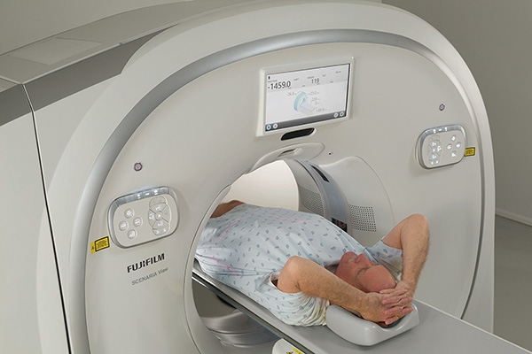
Lateral Shift Table, ± 10cm.

IntelliCenter Auto-Lateral Shift Table is the only standard table that can shift up to ± 10cm laterally (20cm total range) to:

  • Assist patient positioning
  • Reduce dose*
  • Improve spatial resolution

Multi-Bowtie Filters.

Multi-Bowtie filter size selection, based on anatomy to be imaged, reduces radiation dose* in areas outside the filter’s field of view. When used together with the auto lateral shift table, off-center anatomy can be brought to iso-center and the smallest bow-tie filter utilized.

Intelli EC Plus.

Fujifilm’s proprietary 3D mA modulation technology, Intelli EC, automatically modulates mA to lower individual patient dose levels depending on patient anatomy and size and considering the level of iterative reconstruction selected. Intelli EC Plus can be set to provide real-time control of mA, to achieve a constant Noise Standard-Deviation in the resulting images.

Higher Data Sampling Rate.

SCENARIA View’s higher data sampling rate provides faster anatomic coverage without sacrificing image quality and reduces motion artifacts.

Wide 80cm Gantry Aperture.

A more open 80cm gantry aperture spaciously accommodates the most challenging patients.

550 lbs. Table Weight Capacity.

Higher standard weight capacity to accommodate larger patients up to 250 kg (550 lbs.).

Reduced Dose for Cardiac CTA.

ECG Dose Modulation reduces mA during the cardiac phases of a retrospectively gated exam that are not intended for image reconstruction.

IPV – Iterative Reconstruction.

IPV is Fujifilm’s Next-generation “vision modeled” iterative processing that provides dose reduction and overcomes a conventional IR weakness by IPV maintaining normal image texture that better matches the image texture appearance of Filtered Back Projection (FBP). Utilizing IPV vs. conventional FBP reconstruction can provide*:

  • Dose reduction up to 83% at the same image quality (low contrast detectability)
  • Image noise reduction up to 90% at the same dose
  • Noise reduction rate control according to the IPV level used
  • Low contrast detectability improvement by up to 2.0X (100%) at the same dose
  • Natural image texture appearance (the shape of noise power spectrum) similar to that of FBP, even if using higher IPV strength level

Reduced kV.

Four settings support lower kV and dose based on individual patient size. Lower kV provides lower dose for small and pediatric patients.

  • 80kV
  • 100kV
  • 120kV
  • 140kV


Axial Snap-Shot Imaging.

Pulses the X-ray “on” only during a phase of the cardiac cycle using prospectively gated step-and-shoot cardiac scanning – reducing radiation dose.

Dose Reporting and Awareness.

SCENARIA View provides both DICOM Dose Structured Report and a Simple Dose Report to satisfy the full range of your IT and physician reading dose reporting needs. And, SCENARIA View includes Dose Check, compliant with NEMA XR-25, to monitor for exam doses that may exceed notice or alert levels.


3D Auto Positioning Camera

3D Camera

Powered by Fujifilm’s REiLI artificial intelligence (AI), our 3D camera technology ensures optimal positioning of the patient within the gantry to acquire the scanogram images. The ceiling-mounted 3D camera recognizes the entire body anatomy. Once the target region is selected, the patient table can move with one button to the precise start and height position and automatically adjust laterally to place the anatomy in the gantry Isocenter. The console displays the 3D camera image, enabling the technologist to plan the CT scanogram with precision and efficiency.

AutoPose

The 3D auto positioning camera combined with the feature called AutoPose enhances scan workflow and dramatically reduces examination time by automating and shortening the operator’s procedure steps. AutoPose, using an automated algorithm, automatically suggests the scan range and dimension based on protocol and patient’s size observed during the scanogram (Scout) scan. The scan plan (box) is automatically positioned on the scanogram for ease and consistent results while minimizing the time for patient setup.

Technology

Rotation times (seconds) 0.35, 0.4, 0.5, 0.75, 1.0, 2.0
Data collection speed 2880 views per second
Minimum Slice thickness (mm) 0.625
FOV (mm) 20-500
Max scan range (in/mm) 79/2000
Max scan rotations 100
Volume scan pitch 0.578 to 1.578
Scanogram with real-time display AP & Lateral
Normal scan axial mode
Volume scan helical mode
Shuttle scan mode (option) Axial or Helical
ECG Scan modes (option) Retrospective and Prospective
Gantry aperture 80cm
Gantry tilt +/- 30 degrees
Gantry Display 15 inch LCD,
Scan localizer x, y, z
Type Solid state ceramic
Coverage 4cm with 64 rows
Detector elements 64 x 888
Anode heat capacity 7.5MHU
Anode max. cooling rate 1,386kHU/min
Tube cooling Oil/Air
Focal Spot (mm) 0.7 x 0.8; 1.2 x 1.4
Output 84kW
kVp selection 80, 100, 120, 140kV
mA selection 10-700mA (5mA steps)
Table-top width 18in (45.7cm)
Table weight capacity 550 lbs (250kgs)
Horizontal accuracy ±0.25mm
Vertical travel range as low as 49cm to 97cm
Lateral Shift Motion ±10cm (20cm total range)
Scannable range 200cm
Display single 24-inch LCD widescreen monitor
Display resolution 1920 x 1200
UPS for console and image processor Included
Matrix 512 x 512
Image reconstruction time up to 60 images/second (depending on protocol)
Immediate image preview Preview Recon
CT number range -32,768 to +32,767
  • Simple Dose Report
  • DICOM Dose Structured Report (SR)
  • CT Dose Check (NEMA XR-25)
  • NEMA Smart Dose Compliant (XR-29)
Minimum room size 285 Sq. Ft.
Typical room size 520 Sq. Ft.

Focus Edition

Innovative Focus Edition advantages.

Continuing Fujifilm’s commitment to innovative healthcare, the SCENARIA View Focus Edition CT platform is uniquely designed with performance advantages to better meet the needs of a wide range of patients and providers. The Focus Edition is an outstanding CT for Radiology, Cardiology and Emergency Departments as well as outpatient locations. It leverages Fujifilm’s more powerful “Focus Engine” image processor and operator console, resulting in valuable benefits beyond our standard SCENARIA View CT Platform:

  • 20% faster workflow, for more patient exams per day
  • UI update to the Synapse 3D Viewer on the Operator’s Console
  • Tilted gantry helical scanning feature for faster workflow and improved MPR capability
  • AI 3D camera and AutoPose for patient positioning for scanogram and planning
  • Higher level of cardiac CTA capability with “Cardio StillShot” 3D motion correction (optional)

Cardio StillShot

The SCENARIA View Focus Edition’s optional Cardio StillShot software is revolutionary, with a proprietary algorithm that creates clear, high-quality images for even the most challenging heart rates.

  • 28 msec effective temporal resolution at 0.35 sec gantry rotation speed versus 175 msec native temporal resolution without Cardio StillShot – a 6-fold improvement to freeze cardiac motion
  • Motion correction for the coronary arteries, entire heart and the valves
  • Enables more exams to be performed with Prospective Gating versus Retrospective Gating due to lower patient radiation exposure

Enhanced Technologies

The Focus Edition’s enhanced clinical performance is driven by numerous technological advances that Fujifilm has refined to create an outstanding CT System, including:

  • Intelli IPV – Vision Modeled Iterative Reconstruction
  • HiVision Detector
  • HiMAR Plus – Metal Artifact Reduction
  • Cardio StillShot 3D Motion Correction (optional)
  • Advanced 3D Visualization Capabilities
  • AI 3D Camera and AutoPose
Customer Stories

Nashville General Hospital aimed to enhance their mammography and CT systems to remain competitive against larger local medical facilities. Throughout the selection process, Fujifilm stood out as the right fit.

Nashville General Hospital

Nashville, TN

With a goal to provide the fastest, most productive method of obtaining a diagnosis with the best image quality possible and low dose capabilities, Nashville General Hospital turned to Fujifilm’s Scenaria View CT Scanner.

Nashville General Hospital

Nashville, TN

Fujifilm helps West Feliciana Hospital provide quality health care their local families need. With new imaging equipment, they were able to recruit specialists and incorporate additional diagnostic offerings into their care services.

West Feliciana Hospital

St. Francisville, LA

The Scenaria View CT scanner is advantageous for Massac Memorial Hospital’s rural community. It can accommodate a larger number of patient types with its high weight limit and wide bore, while the lateral shift feature (…)

Massac Memorial Hospital

Metropolis, IL

West Feliciana Hospital is a small and unique Critical Access Hospital that recently purchased two key pieces of imaging equipment from Fujifilm to help provide the best care possible to their community. Hear what they (…)

West Feliciana Hospital

St. Francisville, LA

Brochures

SCENARIA View Product Brochure.
SCENARIA View Product Specifications.

SCENARIA View Product Brochure.

Experience enhanced workflow speed and clear CT image quality across a wide range of applications using 64 slice acquisition and up to 128 slice reconstruction. Access the latest dose awareness, reduction and management features.* Benefit from more open patient access, a compact footprint, new energy efficient technologies and lower cost of ownership with Fujifilm’s SCENARIA View.

*In clinical use, dose saving features may reduce CT patient dose depending on the clinical task, patient size, anatomical location and clinical practices employed. Consultation with a radiologist and physicist are recommended to determine the appropriate dose needed to obtain diagnostic image quality for a particular clinical task.

Download the Brochure

SCENARIA View Product Specifications.

Continuing Fujifilm’s commitment to Innovating Healthcare, the SCENARIA View CT platform is designed with unique practical innovations to better meet the needs of a wide range of patients and providers.

Download the Brochure

Call to action icon

Contact Us

Let us help you find the right product or service that will suit your needs.

Contact Us

*In clinical practice, the use of Intelli IPV may reduce CT patient dose depending on the clinical task, patient size, anatomical location, and clinical practice. Image noise is defined as the standard deviation of pixel values within an area. A consultation with a radiologist and a physicist should be made to determine the appropriate dose to obtain diagnostic image quality for the particular clinical task. Low contrast detectability (LCD) and/or image noise were assessed using reference factory protocols comparing Intelli IPV and FBP. The LCD measured in 0.625 mm slices and tested using the MITA CT IQ Phantom (CCT189, The Phantom Laboratory), using model observer method.