When the COVID-19 pandemic struck beautiful Italy in late February 2020, no region was spared. The country’s outbreak started with three cases, and within five days there were 283—including in Tuscany. An alarming moment was felt across the country a month later, when the civil protection authorities announced that 969 people had died in just 24 hours due to the SARS-CoV-2 virus. It was a very scary time, leading Italy to go under necessary lockdown.
Fortunately, clinicians and administrators at the University Hospital of Pisa had paid close attention to the early cases in Lombardy and Veneto, and how COVID-19 patients were managed there. Entire areas of the University Hospital of Pisa prepared to exclusively house COVID-19 patients—dividing the hospital into COVID zones and non-COVID zones, and organizing corresponding task forces.
Three levels of gravity of COVID-19 were established with corresponding hospitalization options. These included normal hospitalization for those with mild pneumonia, assessment by a pulmonologist for those with moderate pneumonia to plan suitable respiratory support, and finally, intensive care assessment for patients with severe pneumonia with a plan to transfer them to the intensive care unit.
But how was the hospital to determine these levels of gravity? Pulmonary involvement identified by a CT scan proved to be fundamentally important to establish each COVID-19 patients’ level of gravity. The quandary, however, was that even with a CT scan a subjective assessment of the extent of pulmonary involvement is often imprecise and not easily reproduced.
Fortunately, radiologists at the University Hospital of Pisa came up with a way to make the assessment automatically.
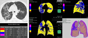
The hospital was using Fujifilm’s Synapse® PACS which supports the use of a powerful image processing tool, Synapse 3D. Using Synapse 3D to evaluate CT scans allowed the clinicians to quickly and reliably identify areas of the lung with increased pixel density —which are typical of COVID-19 pulmonary involvement. This tool, coupled with recommendations from scientific literature, allowed the team to seamlessly create a COVID-19 dataset.

The hospital’s COVID-19 patients could now be classified into specific levels of gravity and, in turn, their management and treatment was apt to be more efficient and effective.
Simply put, the radiologist would process the images using Synapse 3D and on the basis of the ranges assigned, produce output for assessing the density of the lung, and therefore the degree of the patient’s pulmonary involvement.
The process facilitated the hospital’s workflow by providing a rapid assessment. Patients would arrive in the emergency department and receive a swab test followed by a CT scan with a Synapse 3D analysis. This quantitative analysis, along with the help of clinicians’ judgment, provided an objective evaluation so that COVID-19 patients could be triaged according to severity and then managed in the best possible manner.
Back in the early months of 2020, managing COVID-19 patients was no easy task. Thanks to Fujifilm’s Synapse 3D, the job of emergency doctors and pulmonologists at the University Hospital of Pisa was made a little bit easier. And today, the picture for patients and providers is a whole lot brighter.
In the United States, Fujifilm’s Persona CT, a CT solution designed to provide exceptional precision and accuracy for fast decision making, can be integrated and work in parallel with Synapse 3D to provide similar results as the University Hospital of Pisa witnessed. The Persona CT’s open and adaptable equipment, paired with Synapse 3D’s advanced visualization software, make a wider range of scans and treatments possible. These two technologies, working collaboratively, create a truly synergistic care experience.
To learn more about Pisa University Hospital’s use of Synapse 3D and CT to improve patient management during the COVID-19 pandemic, click here.