Search
Just over a month ago, renowned broadcast journalist Katie Couric shocked the world when she revealed she’d privately been battling breast cancer. Receiving the diagnosis after missing her annual mammogram, Couric pleaded with listeners to prioritize the exam that has helped cut the risk of dying from the deadly disease nearly in half.
In this Q&A, The EI Connection spoke with Fujifilm Marketing Manager and former mammography technologist Jeanmarie Rogers, RT(R) (M) (CT), about the progression of mammography technology, the emerging role of artificial intelligence (AI), and how working hand-in-hand with mammographers is enhancing their workflow.
I started my career as a registered radiology technologist at a community hospital performing X-ray exams before venturing into mammography. I started performing mammograms before the industry had dedicated mammography machines and the rules and regulations that we’re accustomed to today. During my time at the hospital, I earned additional certifications in mammography and CAT scan.
I then joined Fujifilm as a workflow specialist when Synapse® PACS was beginning to display digital mammography images. In my current role, I act as the liaison between our customers and development team. My experience as a mammography technologist allows me to understand our customers’ wants and needs, both from a workflow and technical perspective. I’m able to take their feedback to our engineers and help manage the development of new features that help meet our customers’ objectives and goals.
When I first started, our patient volume and image quality were nothing like they are today. Women typically only came in when there was suspicion of a mass or when a lump was found during a physical or self-examination. We would perform two-view mammograms in a regular X-ray room using flexible cassettes to mold under the breast and positioning sponges to hold the breast and the cassette in place.
Our first dedicated mammography machine using film cassettes provided much better image quality, allowed us to perform more positioning views, and gave us the ability to spot-focus on an area of concern better then we could in the past. Digital mammography, with higher-resolution images and better tools than film, really transformed the ability to detect the smallest breast cancers earlier.
Digital mammography streamlined workflow for technologists and radiologists. For technologists, it eliminated the time to perform an exam, stamp the patient’s information on the films, process the films, and hang the current and prior films on the motorized viewbox. Instead, you could have a patient in and out with their images ready for review in less than half the time. For radiologists, it provided immediate access to the current study adjacent to all the patient’s prior studies. This workflow enhancement subsequently allowed facilities to increase the number of patients they could see in a day.
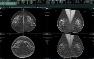
In the past, breast tomosynthesis was only viewable on dedicated mammography workstations, which limited access for referrers and radiologists. Today’s enterprise PACS architecture has radically changed that.
Fujifilm’s PACS reading environment now provides radiologists immediate access to the patient’s entire breast imaging history. Our Synapse PACS PowerJacket, for example, displays all the patient’s current and prior multi-modality breast imaging and reports in a single, all-encompassing view. Our CommonView feature brings in images and reports from other affiliated facilities, as well as from our Synapse VNA, to help ensure the complete patient breast imaging record is accounted for. Our seamless synchronization with mammography tracking systems and electronic health records (EHRs) visual desktops also gives radiologists additional clinical information, all on one workstation. It’s these types of technologies that go beyond just enhancing workflow; they improve diagnostic decision making by presenting the complete patient picture to the radiologists and care teams.
When computer-aided detection (CAD) first entered the mammography space, it would guide radiologists to give potential areas of concern a closer look. At first, radiologists were a little unsure of the technology, but now CAD is a standard tool for diagnosis. The emergence of AI CAD provides radiologists with even more information and tools. Our Synapse AI Platform goes beyond identifying an area of concern by bringing AI directly into the Synapse PACS workflow. Using advanced technology, it triages and prioritizes the studies on the radiologists’ reading worklists based on findings. This type of technology helps providers detect, diagnose, and treat patients sooner and helps reduce the number of false positives.
As a former mammography technologist, I understand our customers’ workflows and challenges. Because of that, I truly take pride in working with them to support their success and achieve their goals through our technology. It’s very important to collaborate with our end-users, as Synapse is designed based on our customers’ direct feedback.
For example, I recently collaborated with several radiologists to get their insight on a new feature we were considering. I shared their insights, suggestions, and enthusiastic feedback with our development team, and we’re now offering that feature in an upcoming release. Sharing feedback in this way helps us continue to advance our mammography product roadmap. It’s interactions like this that help Fujifilm grow.
Knowledge is power; get to know your body through regular self-exams, and prioritize annual mammograms. The earlier you feel something, the higher your recovery and survival rate. With so much technological advancement at our disposal, we’re really at an inflection point. It still amazes me how far we’ve come since I started, and it just keeps getting better.
Want to learn more about Fujifilm’s PACS mammography offering? Click here to book a meeting with our specialists at RSNA booth #1929.