Search
Scoliosis, an abnormal lateral curvature of the spine, is most often diagnosed in children aged 11 and older.1 It can accompany certain neuromuscular conditions like cerebral palsy or muscular dystrophy, but more than 80% of cases are idiopathic – of unknown causes – and likely result from a combination of genetic factors and hormonal dysregulation, among others.2
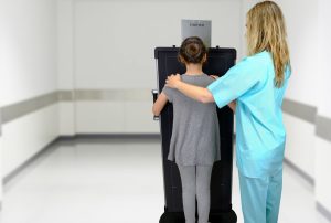
Most scoliosis cases are mild and present no clinical symptoms or pain,3 but some curves can worsen as children grow.4 Severe scoliosis can be associated with adverse health outcomes both in childhood and later in life, including impaired lung and heart function,5,6 emphasizing the importance of timely diagnosis, treatment, and management. Children who have mild scoliosis are therefore monitored closely – typically with routine X-rays – to evaluate curvature development and determine whether a treatment plan is needed and, when in place, effective.
Scoliosis progression can be unpredictable, and patients receive routine screening that can range from 10-25 to as many as 40-50 radiographs over their care history, depending on the severity of the curve.7 Initial diagnosis calls for higher radiation to allow for visualization of bone structure details, whereas follow-up assessments monitor spine curvature and skeletal maturity and can be achieved at lower doses. Nevertheless, repeat radiography using conventional systems has raised concerns about radiation exposure,8
particularly in children and adolescents, where it has been linked to increased cancer risk.9
Although a new, dedicated “ultra-low dose” radiation system – EOS dual planar scanning – exists and is thought to significantly lower the risk of cancer in children (particularly girls),10 it has major disadvantages over conventional systems. The EOS is considerably larger and more expensive, it is slower in image acquisition (and therefore more prone to motion artifacts), and it requires that patients stand or sit by themselves without external help from guardians or staff, making it less suitable for use in younger or developmentally delayed patients.
To overcome these hurdles, a team at Yale School of Medicine created an imaging protocol for follow-up scoliosis X-rays using a modified technique11 that tailors exposure to the patient’s height and weight. In addition, it uses conventional imaging equipment in combination with Fujifilm’s FDR D-EVO GL detector and Virtual Grid anti-scatter image processing software.12 The protocol was validated by comparing images from 40 pediatric patients obtained using either the standard technique or the modified technique. Their results show that the modified technique allows for a 74% reduction in the effective dose, indicating that the incorporation of Fujifilm technology allows conventional radiographic equipment to achieve the very impressive low dose levels of the EOS system.
This new lower-dose protocol is currently used at the researchers’ institution for all follow up imaging of scoliosis patients, further attesting to its impact and perceived efficacy among medical providers. A commentary published in a recent issue of Pediatric Radiology highlights the study’s findings, indicating that their use of height- and weight optimization and replacement of physical anti-scatter grids with denoising software like Fujifilm’s Virtual Grid are achievable changes that can help reduce radiation exposure not only in scoliosis examinations but also in other examinations.13
The full study conducted by the Yale School of Medicine may be viewed and purchased from the Pediatric Radiology journal’s website here. To learn more about the long-length single exposure detector used in the study or Fujifilm’s latest lightweight, wireless model, please visit the product pages for the FDR D-EVO GL and FDR D-EVO III G80i.
REFERENCES
1 Dunn J, Henrikson NB, Morrison CC, Blasi PR, Nguyen M, Lin JS. 2018. Screening for adolescent idiopathic scoliosis: Evidence report and systematic review for the US Preventive Services Task Force. JAMA 319(2):173-187, PMID: 29318283.
2 Kikanloo SR, Tarpada SP, Cho W. 2019. Etiology of adolescent idiopathic scoliosis: A literature review. Asian Spine J 13(3):519-526, PMID: 30744305.
3 Weiss HR, Karavidas N, Moramarco M, Moramarco K. 2016. Long-term effects of untreated adolescent idiopathic scoliosis: A review of the literature. Asian Spine J 10(6):1163-1169, PMID: 27994795.
4 Bunnell WP. 1986. The natural history of idiopathic scoliosis before skeletal maturity. Spine (Phila Pa 1976) 11(8):773-6, PMID: 3810290.
5 Weinstein SL, Zavala DC, Ponseti IV. 1981. Idiopathic scoliosis: long-term follow-up and prognosis in untreated patients. J Bone Joint Surg Am 63(5):702-12, PMID: 6453874.
6 Huh S, Eun LY, Kim NK, Jung JW, Choi JY, Kim HS. 2015. Cardiopulmonary function and scoliosis severity in idiopathic scoliosis children. Korean J Pediatr 58(6):218-23, PMID: 26213550.
7 Oakley PA, Navid Ehsani N, Harrison DE. 2020. 5 Reasons Why scoliosis X-rays are not harmful. Dose Response 18(3):1559325820957797, PMID: 32963506.
8 Oakley PA, Ehsani NN, Harrison DE. 2019. The scoliosis quandary: are radiation exposures from repeated X-rays harmful? Dose Response 17(2):1559325819852810, PMID: 31217755.
9 Doody MM, Lonstein JE, Stovall M, Hacker DG, Luckyanov N, Land CE. 2000. Breast cancer mortality after diagnostic radiography: findings from the U.S. Scoliosis Cohort Study. Spine (Phila Pa 1976) 25(16):2052-63, PMID: 10954636.
10 Rose LD, Williams R, Ajayi B, Abdalla M, Bernard J, Bishop T, Papadakos N, Lui DF. 2023. Reducing radiation exposure and cancer risk for children with scoliosis: EOS the new gold standard. Spine Deform 11(4):847-851, PMID: 36947393.
11 Hoerner M, Grizzard K, Moroz J. 2022. Method of determining technique from weight and height to achieve targeted detector exposures in portable chest and abdominal digital radiography. J Appl Clin Med Phys 23(7):e13582, PMID: 35262265.
12 Al-Dasuqi K, Taylor E, Ehrlich L, Cooperman D, Socci A, Tuason D, Hoerner M, Staib L, Silva CT. 2024. Performance and reliability assessment of a lower dose, task-based scoliosis radiography protocol in pediatric patients. Pediatr Radiol 54(1):146-153, PMID: 38010426.
13 Don S, Moore QT, Hensley P. 2024. Commentary: Achieving ALARA in scoliosis examinations by using body measurements to set techniques and eliminating physical anti-scatter grids. Pediatr Radiol 54(1):154-156, PMID: 38047922.