Synapse® 2D Fusion fuses multimodality and multidimensional data for purposes such as breast or prostate MR or police science applications. It superimposes two 2D / 3D images of the same/different modality.
Synapse® 2D Fusion fuses multimodality and multidimensional data for purposes such as breast or prostate MR or police science applications. It superimposes two 2D / 3D images of the same/different modality.
 Synapse® 2D Viewer provides simple viewing of multimodality images in a single application. It is also embedded as the simple CD/DVD viewer.
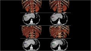
 Synapse® Compositor helps with complex surgical interventions. It enables fusion of up to five series in the same space and displays the volume rendering of combined images. Applications include display of CT images of the liver exposed at multiple time phases, CT images of bone fused with MR images of soft tissue, and MRA images of artery with phase contrast of vein.
 Synapse® 3D Viewer allows orthogonal, oblique, and endoscopic analysis of CT, MR, NM, and PT data.
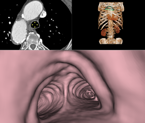 Synapse® 3D Viewer with VE allows a fly-through with contrast-enhanced vessels or hollow structures.
 Synapse® 3D Comparison enables side-by-side comparison and synchronization of multiple 3D data.
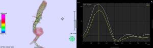 Synapse® 4D Flow calculates the blood flow volume, velocity, and vector of a designated ROI and overlays them onto the extracted region. Flow volume and velocity measurements are also displayed on an analysis table using the time-intensity curve.
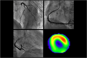
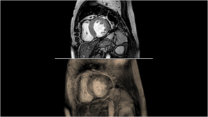 Synapse® 4D Viewer enables CT and MR multiphasic data to be viewed in cine mode. It is useful for viewing cardiac CT/MR. In cine play, 2D cross-sections and 3D images are synchronized.
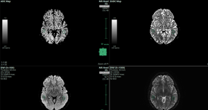 Synapse® ADC Viewer calculates apparent diffusion coefficients from information on signal values of diffusion-weighted images (DWI) collected with MR. The software displays and measures ADC and eADC values in ROIs.
 Synapse® Combination allows stitching of multiple series into a single view.
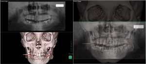
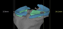
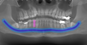 Synapse® Dental MPR creates panoramic and cross-sectional images along teeth and alveolar bones from CT images to assist with dental implant planning.
 Synapse® Dual Energy creates a virtual mono image from two different kV CT series, two or three material decomposition images, or one virtual non-contrast image.
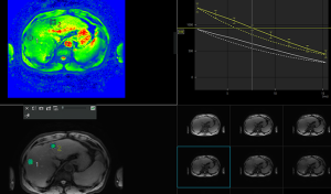
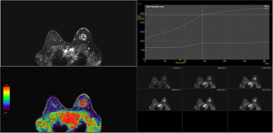 Synapse® Dynamic Data enables clinical assessment of images over time, including breast and prostate MR and dynamic PT analysis. It displays individual parameter images or time-intensity/time-activity curves of slices of multiphase data.
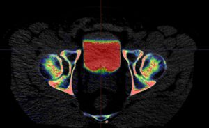
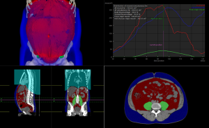 Synapse® Fat Analysis 2D and 3D calculates areas and volumes of subcutaneous and visceral fat in multiplanes (3D). The psoas muscle is automatically extracted and volumes are calculated.
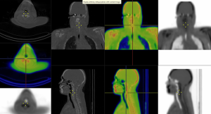 Synapse® Fusion Viewer enables the display of images fused from two different modalities (CT/PT, CT/MR, CT/SPECT, etc) or images of the same modality with different exposure methods, exposure times, etc.
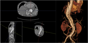
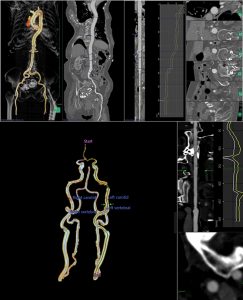 Synapse® General, Aortic, and Carotid Curved Planar Reformat (CPR) are designed to create CPR images for clinical analysis of blood vessels, including stenosis measurements, stent graft planning, and calcification analysis.
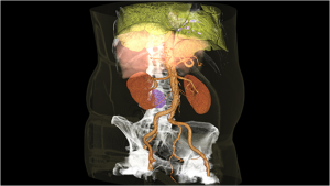
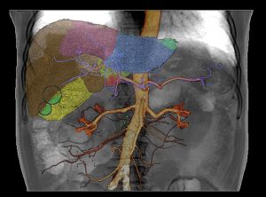 Synapse® Interventional Radiology (IVR) Simulator provides specialized tools to guide preoperative IVR planning and offers preliminary simulation by extracting regions and paths to the target. Use Couinaud segmentation to partition the liver into eight sections for embolization planning.
Synapse® Knee Joint Analysis performs automatic segmentations of bones, meniscus, and knee joint cartilage for various analyses.
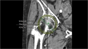 Synapse® MPR Reformat creates a plane along a straight line or in the shape of a fan on 2D images and prints or saves the plane as a new image. It links directly to the 3D Viewer for additional analysis.
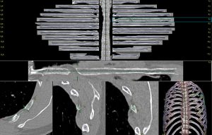 Synapse® Rib Viewer automatically extracts and labels the spine and ribs. Results are displayed in 3D volume and curved planar reformat (CPR) images.
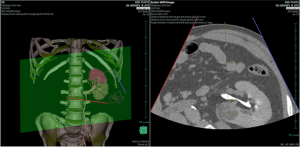 Synapse® Sector MPR simulates ultrasound examinations, particularly for aspiration and biopsy planning, and is ideal for probe- and centesis-placement visualization.
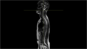
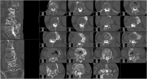 Synapse® Slicer is used for reconstructing spine data through various angles to assist with complex spinal analysis, such as scoliosis evaluation.
 Synapse® Surface Viewer creates STL files for 3D printing* and interactive PDFs for reports or presentations.
 Synapse® Tx Map provides calculations derived from MR signal values. Clinical utility of T2 assessment includes cartilage and collagen analysis to determine iron deposits and distribution.
