ELUXEO Vision

Revolutionizing surgical visualization
with real-time tissue oxygen saturation (StO2) imaging

Gain the clarity of ELUXEO Vision

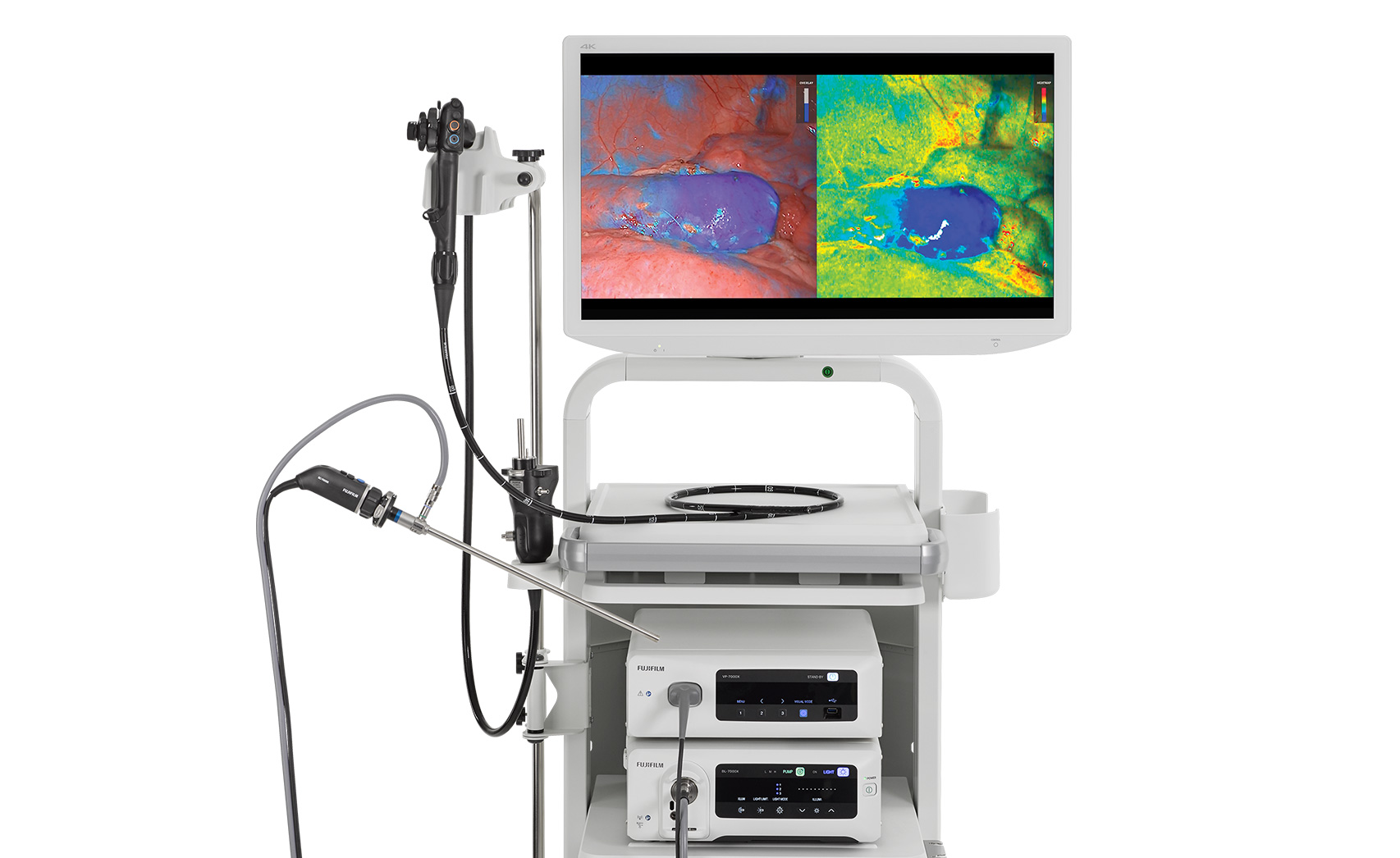
ELUXEO Vision StO2 imaging harnesses the power of data to support informed and confident intraoperative decision-making. It is the only in-market solution that enables surgeons to continuously visualize, quantify, map, and monitor tissue blood oxygen saturation using an endoscope or laparoscope in real-time without requiring dye.

ELUXEO Vision highlights:

  • Quantifies tissue oxygen saturation levels in numerical values
  • Provides a holistic view of the anatomy in multiple visualization modes
  • Continuously monitors serosal and mucosal tissue oxygen saturation
  • Facilitates efficient and repeated tissue viability evaluation
  • Available as an upgrade to the ELUXEO® Surgical System and ELUXEO® Endoscopic Imaging System

Transform how you evaluate tissue viability in the OR.
Make data-backed decisions with ELUXEO Vision.

CONNECT WITH US


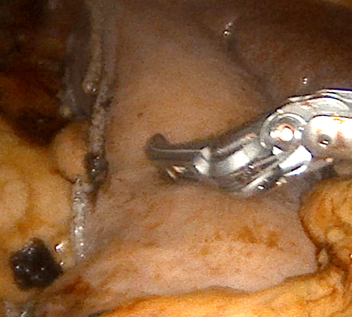
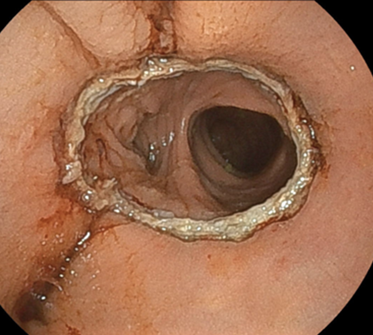
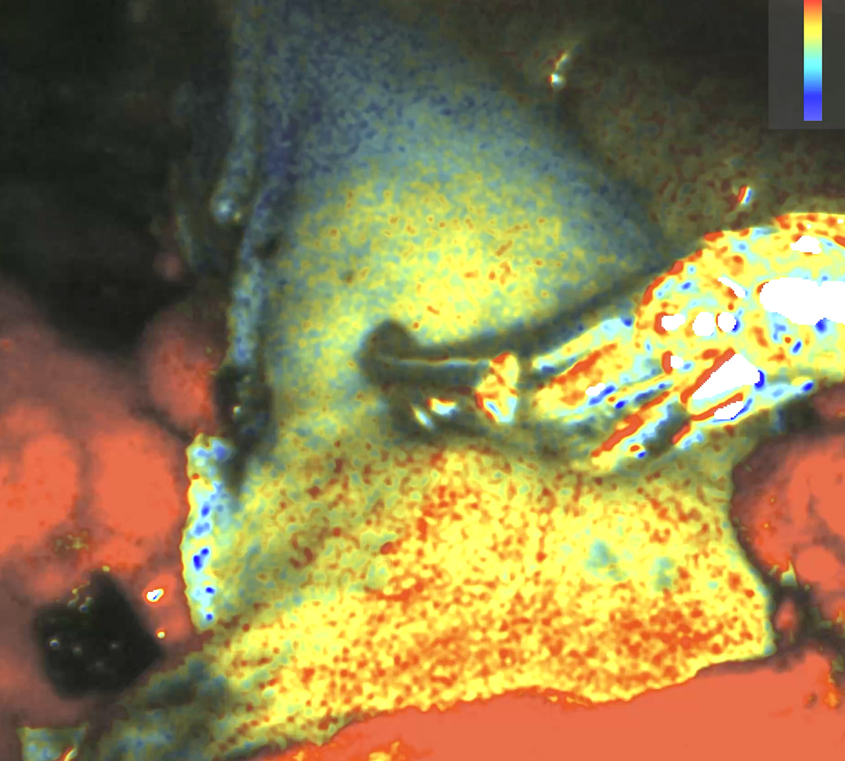
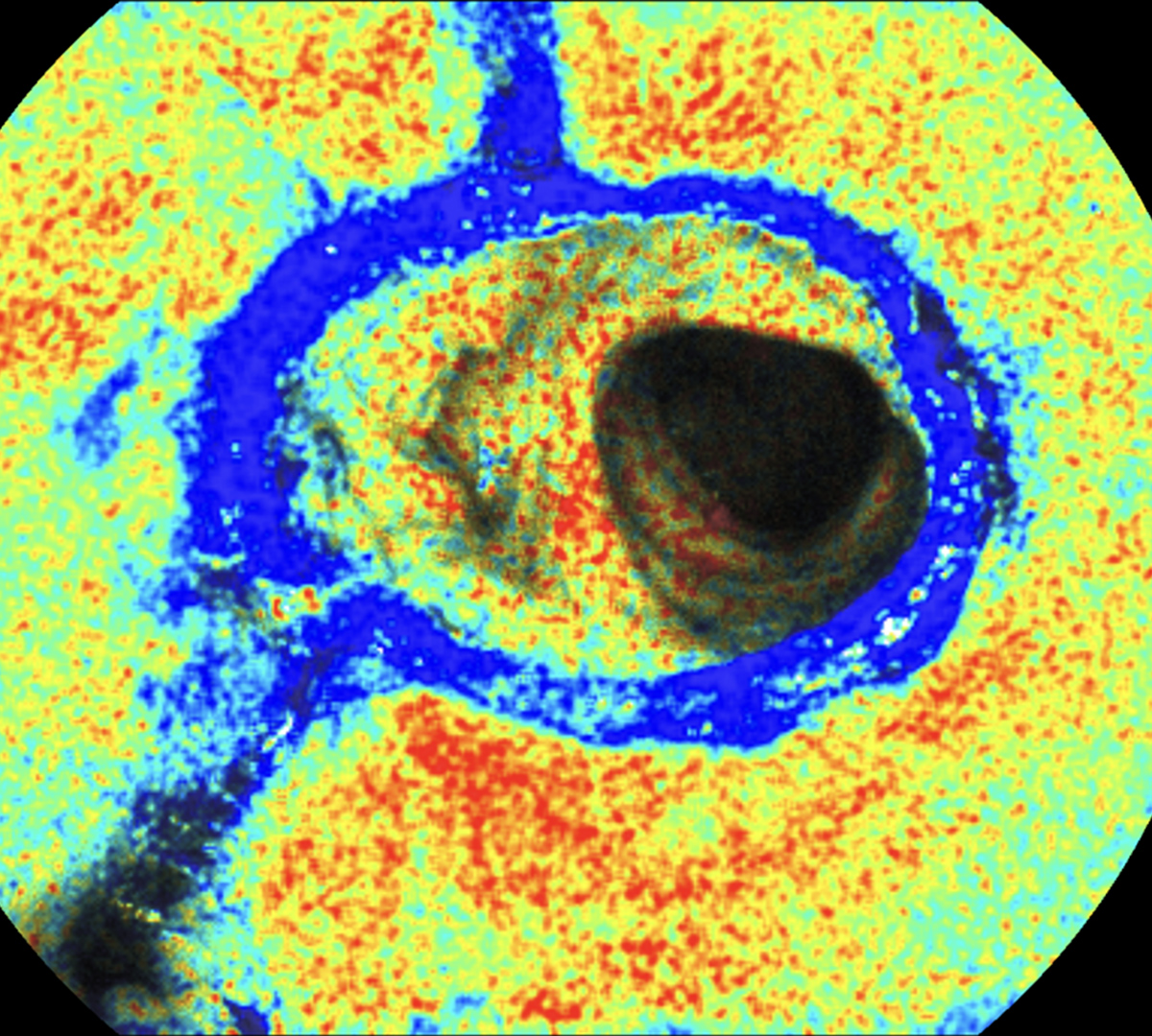
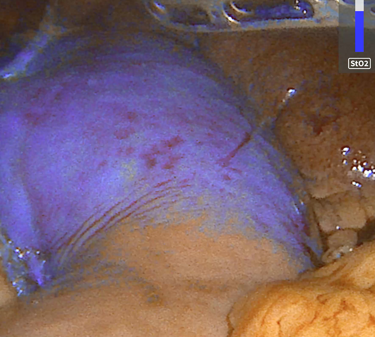
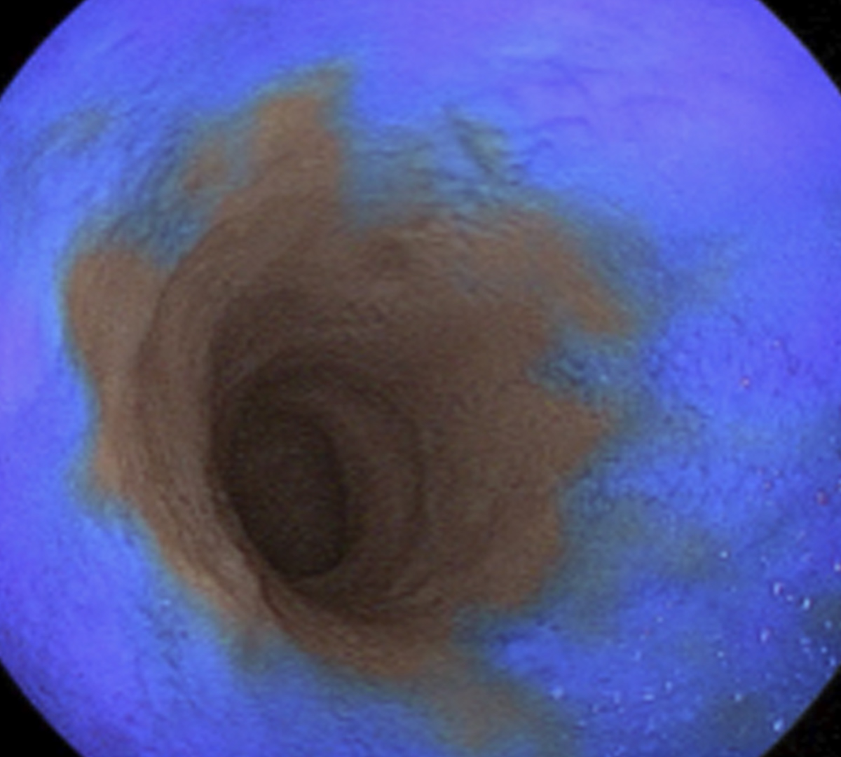
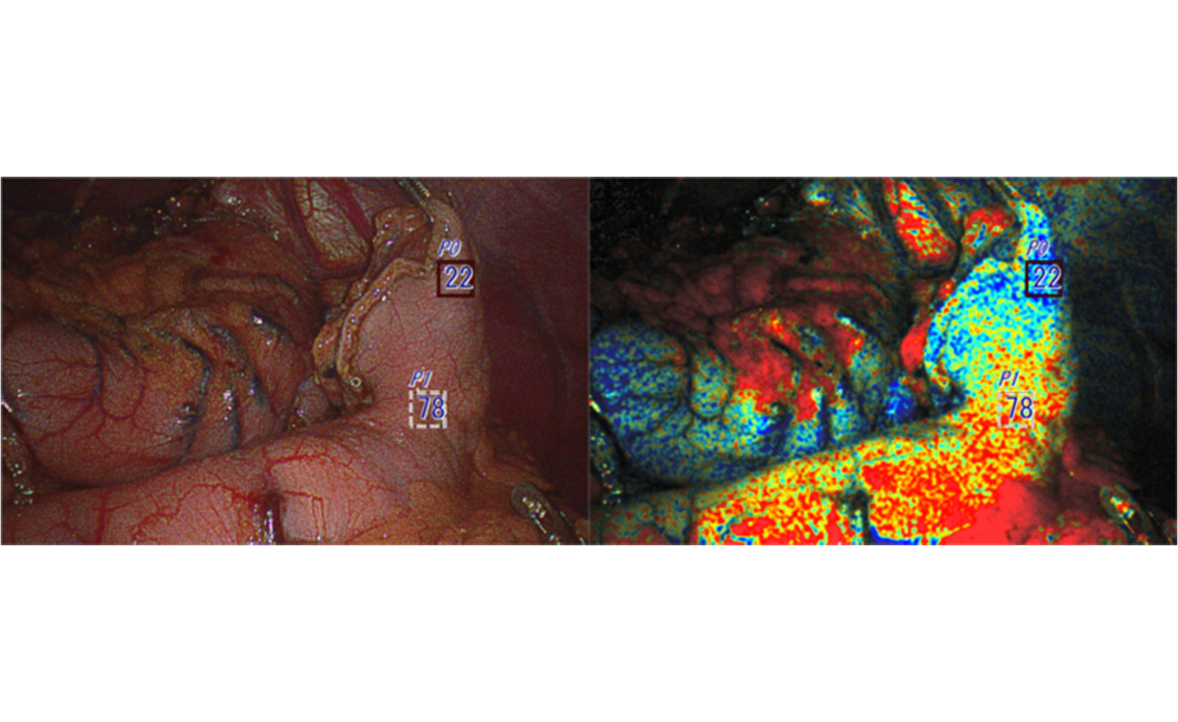
ELUXEO Vision image comparison with White Light, Heatmap, and Overlay modes Laparoscopic Endoluminal
White Light Imaging (WLI)
Brilliantly clear LED multi-light technology provides unparalleled image quality and elevated clarity.
ELUXEO Vision Heatmap
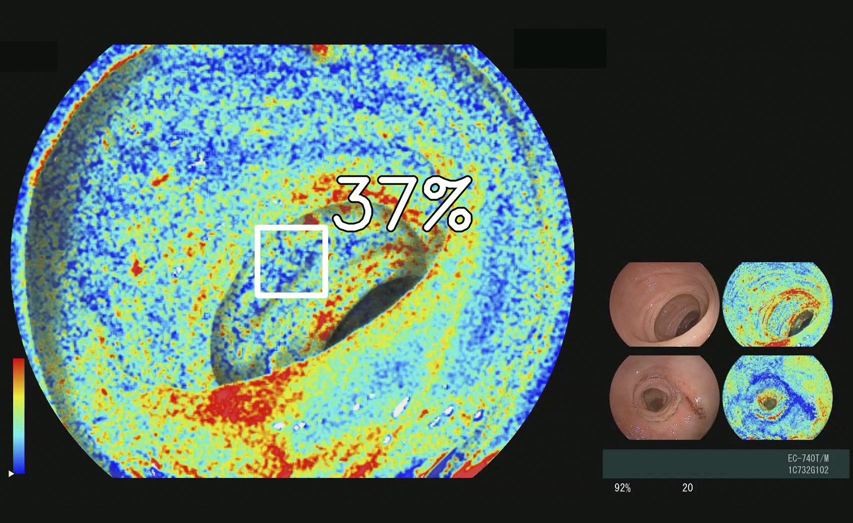
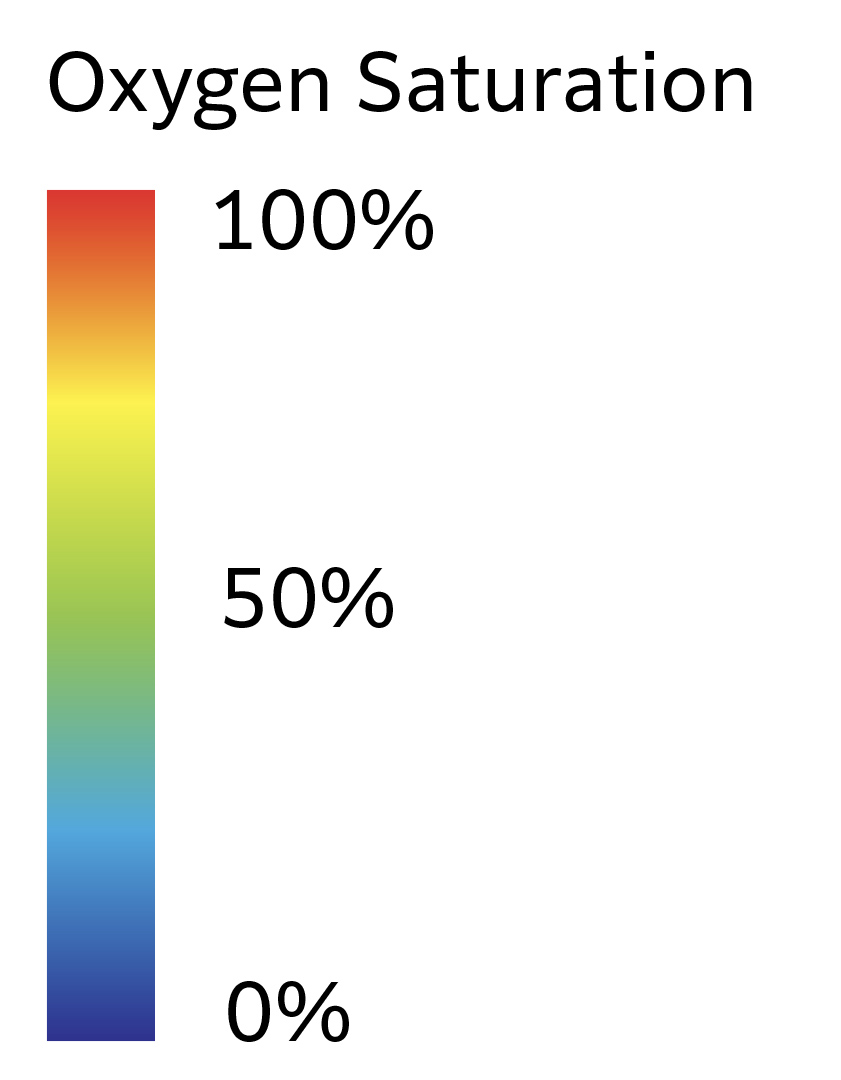
Heatmap mode displays patient anatomy using a color-coded heatmap and key. ELUXEO Vision delivers quantitative data in numeric percentage readings from 0% to 100% and qualitative data as color gradients that correspond to oxygen saturation levels.
ELUXEO Vision Overlay Mode
Overlay mode allows the user to set a threshold, adjustable by 5% increments, which will highlight all tissue in bright blue that is below the preferred oxygen saturation level.

Real-time physiological analytics to aid informed, data-driven decisions

Anastomotic leaks are among the most feared post-surgical complications following any type of gastrointestinal anastomosis,1 including foregut, colorectal, bariatric, and thoracic.

Near-infrared imaging (NIR) with indocyanine green (ICG) is the most used technology for tissue perfusion assessment. However, studies on its utility in intraoperative decision-making have shown varying results.2

Experts call for technological improvements that provide real-time visualization with standardized, quantitative parameters.2-4

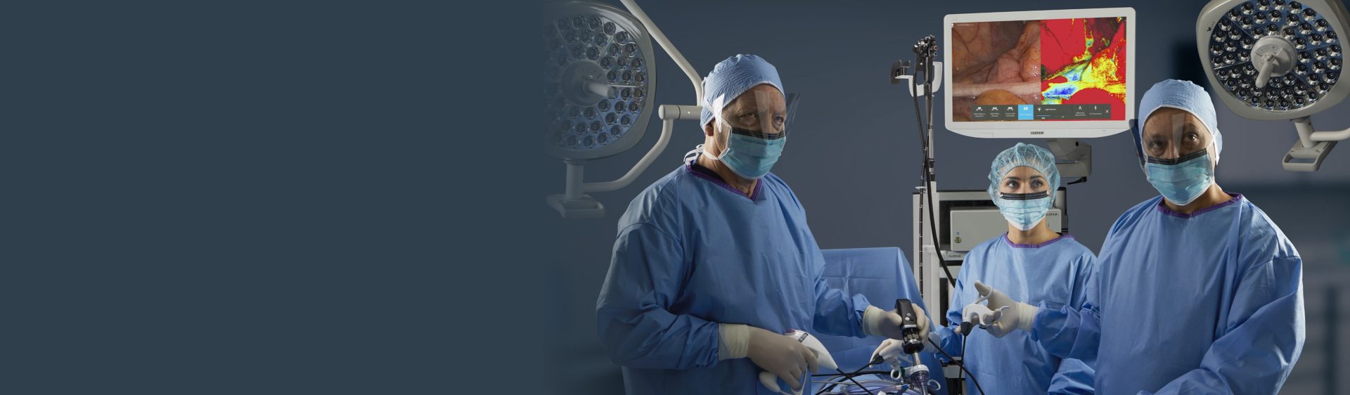
ELUXEO Vision answers the call for innovation in surgical visualization.

Find out how to bring this FDA-designated “Breakthrough Device” to your OR.

CONTACT US

“It [ELUXEO Vision] gives you an absolute saturation without the dyes, as opposed to ICG where it gives you relative oxygen perfusion relative to your standard point.”5

From the study Real time evaluation of tissue oxygenation with Fujifilm’s ELUXEO Vision Surgery System: Addressing clinical challenges in colorectal surgery and beyond

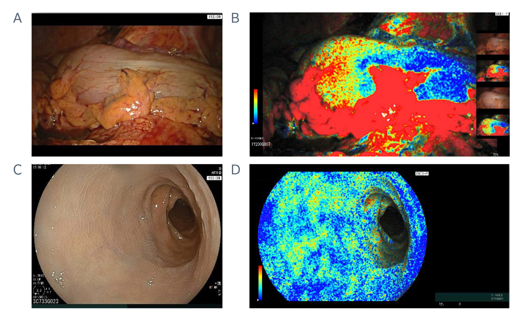
Img: Serosal images (top) using a laparoscope with White Light Imaging (A) and ELUXEO Vision StO2 imaging (B). Mucosal images of the same patient taken with a flexible endoscope with the White Light Imaging (C) and ELUXEO Vision StO2 imaging (D). 5

Flexibility and efficiency for your OR

Offered as an add-on capability to the ELUXEO Surgical System and ELUXEO Endoscopic Imaging System, ELUXEO Vision enables surgeons across specialties to work together from the same tower.

Visualization of StO2 in a patient’s GI tract can assist physicians in identifying tissue ischemia that potentially leads to complications such as anastomosis leaks.

Published studies on anastomotic leak rates for endoscopic thoracic surgery, particularly esophagectomy, report a range of 10% to 30%.6

“OSI [oxygen saturation imaging] can measure gastric conduit perfusion both qualitatively and quantitatively. OSI may be used without delay between steps and may be an alternative to ICG dye perfusion to measure gastric conduit perfusion.”7

From the study 373. novel oxygen saturation imaging to evaluate gastric conduit perfusion during esophagectomy

“The technology allows for continuous, repeatable measurements without the limitations of dye saturation, and it is effective for both laparoscopic and endoscopic visualization.”8

From the study Feasibility of intraoperative tissue oxygen saturation imaging using OXEI technology during robotic esophagectomy: a case series

ELUXEO Vision with flexible scopes

Add ELUXEO Vision to your ELUXEO Endoscopic Imaging System tower to experience more capabilities and efficiencies for your OR. Your OR footprint stays the same, but your reach grows exponentially.

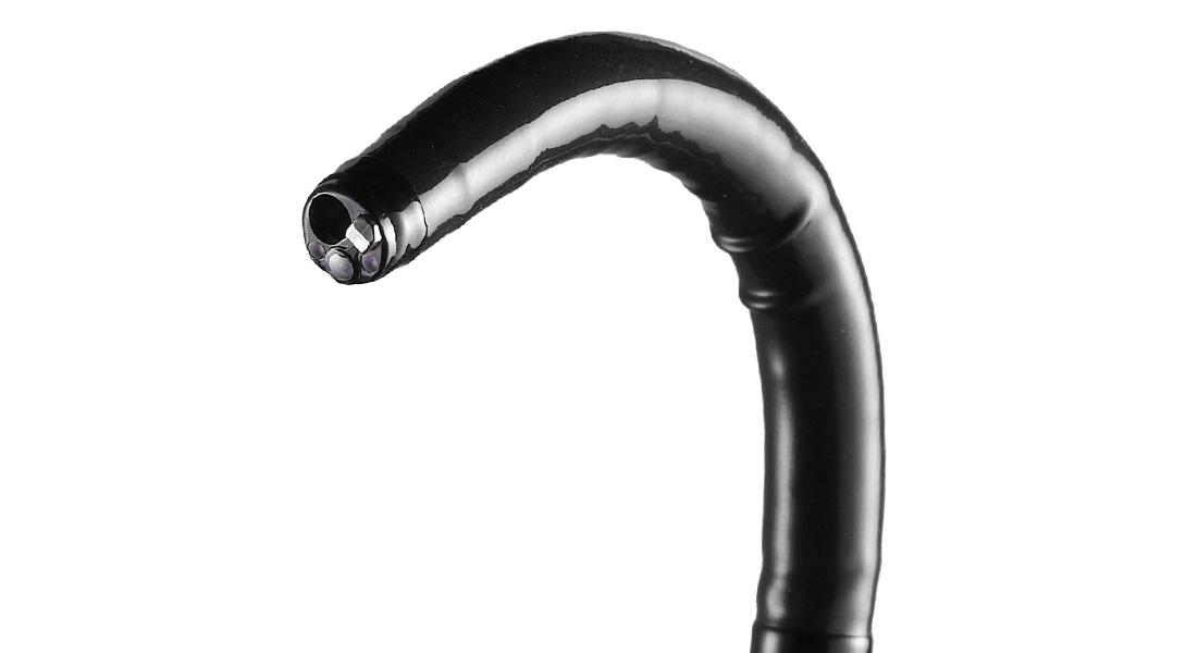
Fujifilm ultra-slim and dual channel flexible scope options support a variety of applications where surgeons can extend StO2 imaging technology to advance complex surgery in the upper and lower GI, foregut, and colon.

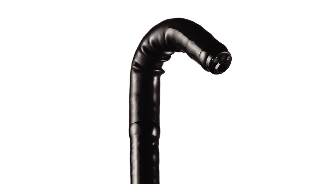
EG-740N Ultra-Slim Gastroscope

  • Slim design supports soft transnasal insertion
  • Large working channel of 2.4mm to allow for superior suction
  • 210° upwards bending radius provides excellent maneuverability and observation
  • Ultra-slim insertion tube, adaptive bending, and advanced force transmission capabilities to help improve insertion and navigation

EC-740T/L Ultra-Slim Colonoscope

  • Smart Bend technology with 210° retroflexion at the distal end, enabling access behind folds for improved detection and enhanced angulation
  • A water jet and a large working channel of 3.2mm in an ultra-slim design
  • Ergonomic G7 control portion designed for ease of movement to help reduce fatigue

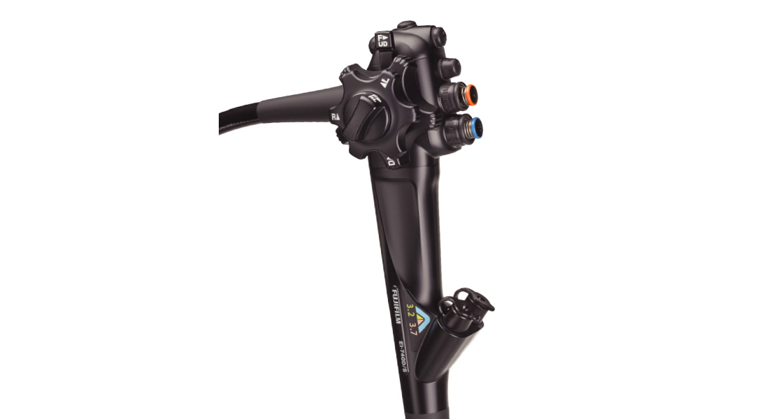
EI-740D/S Dual Channel Endoscope

The first dual channel endoscope FDA cleared for use in both upper and lower gastrointestinal applications and thus broadens the capabilities of gastroenterologists across a wide range of therapeutic procedures. Dual channels of 3.7mm and 3.2mm allow for a broad range of devices for use with the endoscope, including Fujifilm’s TRACMOTION device for ESD, ClutchCutter® and FlushKnife.

ELUXEO Vision with rigid scopes

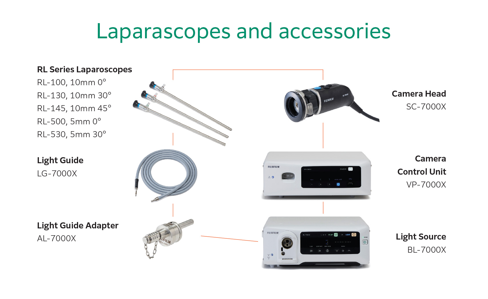
Offered as a state-of-the-art surgical visualization system with 4K or UHD monitors and output, ELUXEO Vision operates as part of the ELUXEO Surgical System, and can be leveraged in conjunction with select Fujifilm rigid scopes to evaluate tissue viability on the serosal side of the GI tract continuously in real-time without dye.

The ELUXEO Surgical System includes the VP-7000X 4K camera control processor, BL-7000X light source, SC-7000X camera head, AL-7000X light guide adapter, and RL series laparoscopes.

Call to action icon

Get To Know Us Better

Let us help you find the right product or service that will suit your needs.

Get in Touch

References

  1. Chadi SA, Fingerhut A, Berho M, et al. Emerging trends in the etiology, prevention, and treatment of gastrointestinal anastomotic leakage. J Gastrointest Surg. 2016;20(12):2035-2051. doi:10.1007/ s11605-016-3255-3
  2. Pollmann L, Juratli M, Roushansarai N, Pascher A, Hölzen JP. Quantification of indocyanine green fluorescence imaging in general, visceral and transplant surgery. J Clin Med. 2023;12(10):3550. doi:10.3390/jcm12103550
  3. Preziosi A, Cirelli C, Waterhouse D, Privitera L, De Coppi P, Giuliani S. State of the art medical devices for fluorescence-guided surgery (FGS): technical review and future developments. Surg Endosc. 2024;38(11):6227-6236. doi:10.1007/s00464-024-11236-5
  4. Larsen PO, Nerup N, Andersen J, Dohrn N, Klein MF, et al. 2023. Anastomotic perfusion assessment with indocyanine green in robot-assisted low-anterior resection, a multicenter study of interobserver variation. Surg Endosc. 2023; 37:3602-3609. doi.org/10.1007/s00464-022-09819-1
  5. Lee SW. Real time evaluation of tissue oxygenation with Fujifilm’s ELUXEO Vision Surgery System: Addressing clinical challenges in colorectal surgery and beyond. Symposium presented at: American Society of Colon & Rectal Surgeons (ASCRS); May 12, 2025; San Diego, CA.
  6. Scognamiglio P, Reeh M, Melling N, et al. Management of intra-thoracic anastomotic leakages after esophagectomy: updated systematic review and meta-analysis of endoscopic vacuum therapy versus stenting. BMC Surg. 2022;22(1):309. doi:10.1186/s12893-022-01764-z
  7. Ferrel M, Ishikawa Y, Chang A, Reddy R. 373. novel oxygen saturation imaging to evaluate gastric conduit perfusion during esophagectomy. Diseases of the Esophagus. 2025;38 (Supplement_1). doi.org/10.1093/dote/doaf061.226
  8. Fortich S, Den J, Thomas M, Petrov, R. Feasibility of intraoperative tissue oxygen saturation imaging using OXEI technology during robotic esophagectomy: a case series. Innovations: technology and techniques in cardiothoracic and vascular surgery. Published online February 3, 2026. doi.org/10.1177/15569845251401204