Search
In the MRI suite at St. Joseph’s Candler Hospital in Savannah, Ga., the numbers add up to a quantifiable success story. Monthly scan counts are up by as many as 150 per month. Business has increased 16.5 percent over the previous year. And back-outs—instances where fearful patients decline to go through with a scan—have plummeted from as many as two per day to just two over the past six months.
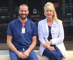
What accounts for the sterling performance? Imaging manager Cheryl Rawlings and registered CT/MR technologist Jonathan Nelson answer by pointing to the confluence of three key contributing factors: the entire imaging team’s unfailing dedication to genuinely compassionate patient care, MR technologists’ beefed-up technical skill sets and, most dramatically, the April 2016 replacement of a legacy MR system with Echelon Oval.
The latter is Fujifilm’s large-bore scanner that delivers crisp whole-body images and state-of-the-art clinical capabilities at 1.5T. The system incorporates workflow features to streamline all relevant tasks and processes, from patient prep to acquisition planning and from image acquisition to post-processing.
The installation also included a suite makeover complete with ambient lighting and patient-sensitive air circulation. But, Rawlings and Nelson agree between them, it’s the size of the bore—at 74 cm in bore width, the widest MRI system available—and the uncompromised quality of the images that have gone furthest in making Candler’s MRI capabilities the talk of the town.
“We have a lot of competition for MR, especially from neurology and orthopedic practices in town and freestanding MR facilities in the area,” says Rawlings, adding that Candler’s MR caseload is 75% to 80% outpatient. “Now we’re working to build our vascular business as well, and a big part of why we’re doing so well with referrals is that claustrophobic patients don’t leave and larger patients are accommodated.”
In fact, the Oval’s table can support patients up to 550 pounds. “We had been sending those larger patients to a different site,” says Rawlings, “and the images weren’t anywhere near the quality we’re getting from the Fujifilm.”
When the Candler MR team went shopping for a new system, Fujifilm already had something of a leg up even though the hospital had been true to another imaging vendor for many years. The advantage came in the form of Candler’s high satisfaction with the quality, service and support they had experienced with the Fujifilm Scenaria CT system they installed in 2015.
“Another reason we chose the Echelon Oval was the software. Fujifilm gives you all upgrades as soon as they become available,” says Nelson. “And the applications training and support over the months since the go-live has been outstanding, just as with the CT. We can call Fujifilm applications people directly, or we can call the help desk. Either way, you talk to a person. You don’t get a prompt for automated responses.”
Nelson also likes that Fujifilm has staff dedicated to making sure customers have all the help they need to stay accredited with the American College of Radiology.
When it comes to building volume while warding off leakage of patients to other imaging providers, Rawlings and Nelson suggest, Echelon Oval’s unique coil solutions allow the machine to perform like several scanners in one.
“Because we are in a hospital setting, a lot of the patients can’t do exactly what you need them to do in a cookie-cutter way,” Nelson says. “With the way the integrated coils are configured, there’s leeway to have some creative thinking by the technologists to actually get an exam done.”
He says he’s used the coils to image a painful foot without having to use a dedicated boot coil as well as the shoulder of a 435-pound shoulder patient who didn’t fit in the standard shoulder coil. “I was able to get it done using the integrated coils and the flexible extremity coil,” Nelson says.
The unique array of coils also allows for breast imaging of larger patients, which, Rawlings points out, is another business-builder.
“We’re affiliated with a cancer center, and we have some breast surgeons at our back door,” she says. “So that was one of our main focuses when we compared MRs from different vendors. We were able to go and look at the breast center and see that we could get larger breast patients into this scanner. That was a huge factor for us.”
Focused on patient satisfaction as they are, Rawlings and Nelson are attuned to how their improved MRI service—which sees heavy duty for brain imaging, followed by, roughly in order, spine, body and extremity—reflects on Candler’s reputation for topnotch patient experience.
MRI has stayed on the satisfied side of HCAHPS surveys, Rawlings says, because patients and their physicians are pleased with both the experience and the images.
“The patients don’t have to return or be called back like they used to,” she adds. “The studies are now all diagnostic. We don’t regularly have patients coming back for nondiagnostic MR studies or for movement or anything like that. Overall patient satisfaction scores, including our HCAHPS scores, are better because of the quality we are providing now.”
Nelson concurs and adds that the image quality they’re seeing from the Echelon Oval is of high enough quality that they’re confident to try new things at the request of several radiologists.
These “new things” are additional boosters of volume and reducers of leakage.
“We started doing a dynamic pituitary exam, which was something we couldn’t do before,” he says. “We also started doing some volume imaging of the cervical spine. And I guess you could say that has enhanced the experience for doctors as well as patients and techs.”
Rawlings offers another anecdote to illustrate how unmistakably Echelon Oval has helped Candler grow its MRI business and put a plug in patient leakage.
In the past, she says, Candler had to schedule all prostate MR patients at a sister hospital that has a 3T scanner. After they got the Oval, they went ahead and did one of those prostate MR exams for themselves.
“We got our hand slapped a little bit, but the radiologist looked at the prostates being done here—and now he likes getting them done here rather than there,” Rawlings says. “So by getting the Oval we’ve been able to grab some of the exams back that we had been forfeiting to our sister hospital with the 3T.”
Asked what advice they might give other health systems considering Fujifilm for the first time—especially those with staff members inclined to avoid taking a chance on an unfamiliar vendor—Rawlings and Nelson don’t have to pause to come up with an answer apiece.
“I would go on a site visit where Fujifilm is installed and talk to as many technologists as you can,” Nelson says. “They’re the people who work with this equipment every day. Of course talk to the doctors too, especially the ones who read the studies, but just from my standpoint, anyone who does that site visit is going to find that there really isn’t any risk to going with Fujifilm at all.”
“The support far surpasses that of any other vendor we have ever used,” says Rawlings. “Fujifilm’s applications specialists for MRI have been just as helpful as their applications specialists for CT. They’re always available for questions or guidance or whatever our technologists need. They have formed relationships to where they are now like family.”